Can Young Women Get Screened For Breast Cancer?
Young women can and should consider breast cancer screening. Discover key risk factors, when to start screenings, and proactive health tips for early detection.

Young women can and should consider breast cancer screening. Discover key risk factors, when to start screenings, and proactive health tips for early detection.

Discover how breast cancer is diagnosed after screening. Understand essential steps, tests, and what to expect to make informed decisions about your health.

Discover what an abnormal mammogram means for your breast health. Understand follow-up tests, manage anxiety, and take proactive steps for better outcomes.
Get a free consultation and talk through symptoms, treatment options, and possible care paths with our coordination team.
Get Free Consultation
Mammograms are safe for breast cancer screening. Discover key facts that ease concerns, empower informed decisions, and prioritize your breast health today.

Discover the link between breast pain and cancer risk. Understand causes, symptoms, and what to watch for. Empower your health—get informed today.

Dense breasts can hide breast cancer on mammograms. Discover advanced screening options and proactive steps to enhance early detection and protect your health.

Discover essential breast cancer screening guidelines by age to empower your health decisions. Learn when to start screenings and how personal risk factors influence your schedule.

Discover how breast MRI enhances cancer screening, especially for high-risk women. Understand its benefits, limitations, and prepare effectively for your appointment.

Discover the reliability of breast cancer screening methods and how they impact early detection. Empower yourself with knowledge and take charge of your health today.
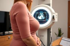
Discover how to detect breast cancer without a lump. Learn key non-lump symptoms and effective screening methods for early diagnosis and better outcomes.
Breast Cancer Screening & Early Detection
Regular screening is the single most powerful tool for lowering breast-cancer mortality because it identifies tumors long before symptoms appear, when treatment is simpler and survival rates exceed 95 percent. Modern programs combine proven imaging tests with risk-stratified follow-up, enabling clinicians to tailor surveillance to each individual’s biology, breast density, and family history. Below is a comprehensive, SEO-optimized guide that explains why screening matters, who should be screened, the technologies involved, and how emerging science is reshaping early detection.
The goals of screening are threefold: detect cancers at Stage 0 or I, reduce the need for aggressive treatments such as mastectomy or chemotherapy, and ultimately prevent deaths. Large population studies show that widespread mammography since the 1990s is responsible for nearly half of the decline in breast-cancer mortality, while advances in therapy account for the rest. Yet disparities persist—screening rates lag in rural areas, underserved urban neighborhoods, and among certain ethnic groups—so expanding equitable access remains critical.
Current Guidelines at a Glance • Women of average risk are advised to begin biennial mammograms no later than age 40, with annual screening an option between 40 and 54. • After age 55, many can transition to mammograms every two years as long as they remain in good overall health and expect to live at least ten more years. • High-risk individuals—defined by BRCA1/2 or other pathogenic variants, a strong family history, previous chest irradiation, or a risk assessment score above 20 percent—should start annual mammography and breast MRI at age 30, sometimes earlier. • Clinical breast exams from a healthcare professional every one to three years supplement imaging in younger women and remain important for those lacking access to mammography. • Self-awareness is encouraged at all ages; knowing your normal breast appearance and promptly reporting changes speeds diagnosis.
Understanding Your Personal Risk Profile Risk calculators combine age, reproductive history, prior biopsies, ancestry, and genetic testing results to categorize people as average, moderate, or high risk. This dictates when to start imaging, how often to screen, and whether to add modalities beyond mammography. For example, women with extremely dense breasts (about 10 percent of the population) benefit from supplemental ultrasound or MRI because dense tissue can mask small lesions on X-ray and independently increases cancer risk.
Key Screening Modalities Mammography: The gold standard uses low-dose X-rays to capture detailed images from two angles. Digital breast tomosynthesis—often called 3-D mammography—creates thin slices that reduce false positives and improve cancer detection in dense breasts. Contrast-enhanced mammography is gaining popularity for its ability to highlight blood-rich tumors.
Breast Ultrasound: Painless and radiation-free, ultrasound excels at distinguishing fluid-filled cysts from solid masses and in clarifying ambiguous mammogram findings. Automated whole-breast ultrasound systems perform a comprehensive sweep, offering a reproducible option for women with dense tissue.
Magnetic Resonance Imaging (MRI): With sensitivity above 90 percent, MRI finds cancers invisible to other tests, making it indispensable for high-risk patients. Dynamic contrast-enhanced sequences highlight abnormal vascular patterns, and recent abbreviated protocols cut scan time to under ten minutes, lowering cost barriers.
Molecular Imaging and Nuclear Medicine: Positron emission mammography (PEM) and molecular breast imaging (MBI) use radiotracers to flag metabolic hot spots. These niche tools help evaluate indeterminate lesions, particularly in dense or reconstructed breasts, though radiation exposure and limited availability restrict routine use.
Emerging Technologies Driving the Future of Early Detection Artificial intelligence algorithms now read mammograms alongside radiologists, flagging subtle distortions and asymmetric densities with accuracy comparable to expert human readers. Early studies show AI triage can reduce workload by 30 percent without missing cancers, allowing radiologists to focus on complex cases.
Liquid biopsies analyze circulating tumor DNA, microRNA, and protein signatures in a simple blood draw. Pilot trials suggest these tests can detect breast cancer months before imaging abnormalities appear. While still investigational, their integration into screening protocols could transform surveillance for high-risk groups.
Microwave and optical imaging, elastography, and low-dose photon-counting CT are also under investigation for their potential to improve specificity or comfort while maintaining sensitivity.
Screening in Special Populations Men account for about one percent of breast cancers, yet delayed diagnosis leads to poorer outcomes. Routine screening is not recommended for average-risk men, but annual mammograms are prudent for those with BRCA2 mutations, Klinefelter syndrome, or significant family history.
Transgender women on estrogen therapy for more than five years accumulate breast tissue and should follow age-appropriate guidelines, adjusted for hormone exposure duration and additional risk factors. Transgender men who have not undergone bilateral mastectomy require screening similar to cisgender women; those with chest reconstruction still retain residual tissue and may need tailored imaging.
Managing Abnormal Results About 10 percent of mammograms lead to callbacks for additional views or ultrasound, and only four to five percent require biopsy. Most biopsies reveal benign conditions, such as fibroadenomas or cysts, underscoring the importance of timely follow-up rather than panic. When cancer is found, catching it early typically means lumpectomy plus radiation, short-course endocrine therapy, or even active surveillance for certain low-grade ductal carcinoma in situ.
Lifestyle Factors That Complement Screening Maintaining a healthy weight, limiting alcohol, engaging in regular physical activity, and choosing balanced nutrition rich in fiber, fruits, and vegetables reinforce the benefits of screening by lowering overall risk. Breastfeeding and minimizing long-term hormone-replacement therapy also confer modest protection.
How to Prepare for Your Screening Appointment Schedule mammograms one week after menstruation when breasts are less tender. Bring previous images or ensure your facility has them for comparison. Avoid deodorant or talcum powder, which can mimic calcifications on X-ray. Communicate breast symptoms, implants, or prior surgeries to the technologist for optimal positioning and exposure settings.
Optimizing Health Equity in Breast-Cancer Screening Barriers such as cost, transportation, language differences, and mistrust can deter timely screening. Mobile mammography units, community-based education, and insurance coverage expansions have boosted participation in many regions, yet continued outreach is vital. Technologies like portable ultrasound and AI-enhanced triage offer scalable solutions for low-resource settings worldwide.
Take-Home Message Consistent, evidence-based screening coupled with individualized risk assessment saves lives by catching breast cancer in its earliest, most treatable stages. Advances in imaging, molecular diagnostics, and artificial intelligence are refining detection accuracy and reducing unnecessary biopsies. By staying informed about guidelines, understanding personal risk, and accessing the right screening tools at the right intervals, every individual can take proactive control of their breast health.